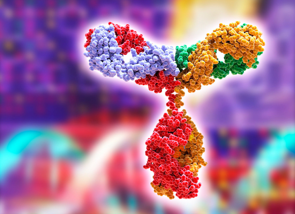
 Immunoglobulin is protein that play a critical role in our immune system. Each class of immunoglobulin has its own chemical structure, biological features, and target specificity. Immunoglobulin is used in research and therapeutic applications to treat a wide range of diseases. It can be obtained from human plasma. Antibodies are Y-shaped proteins made in great abundance by the immune system to help eliminate disease-causing microbes, such as viruses and bacteria. They work by recognizing and sticking to a specific protein on the surface of these organisms, called an antigen. They are made by B cells and are found in the blood, bone marrow, spleen and lymph nodes. They are the most common antibodies and form the basis of the immune defenses against harmful organisms and toxins. During B cell proliferation, they undergo somatic hypermutation (SHM), a process that results in approximately one nucleotide change per variable gene, every time the cell divides. The mutations allow the antibody genes to evolve slightly from their original amino acid sequence, thereby allowing the production of more sensitive and specific antibodies. The two heavy chains and two light chains of an antibody form a polypeptide molecule that is held together by covalent disulfide bonds. They are separated by a region called the constant region, which contains the binding site that is optimized to latch on to a particular foreign protein, also known as an epitope. Immunoglobulin replacement therapy can help people with primary immunodeficiency disease (PIDD) or inborn errors of immunity (IEI). It gives them antibodies that they don't have. It can also help people with autoimmune diseases, like lupus or myositis. It helps their bodies to make more red blood cells to carry oxygen and fight infection. The liquid part of blood, called plasma, is separated out from red blood cells and used to make the Immunoglobulin products. The plasma is collected from large numbers of healthy blood donors who have completed a medical questionnaire and been carefully screened for infection. IGRT is administered via intravenous or subcutaneous routes and is the standard-of-care treatment for patients with PIDD who have impaired antibody production and function. Immunotherapy is a form of cancer treatment that uses the immune system to fight the disease. It usually results in fewer short-term side effects than chemotherapy. There are several types of immunotherapy, including vaccines that stimulate the immune system in case of cancer and targeted drugs that attack specific proteins on cancer cells. These medicines can be used to treat many different kinds of cancer, including leukemia, lymphoma, breast and prostate cancers, and lung and bladder cancers. These treatments can also help the immune system respond to cancer that has already spread to other parts of the body. Research shows that people whose tumors contain a type of cell called tumor-infiltrating lymphocytes have better outcomes when treated with immunotherapy than those whose tumors don’t have TILs. Other types of immunotherapy include checkpoint inhibitors and monoclonal antibodies. These are produced in laboratories and use antigens to trigger immune cells to destroy specific cancer cells. Immunosuppression is the reduction of the body's immune system strength. It can be caused by a disease or drug and may also occur as part of treatment for certain medical conditions. People who have a weakened immune system can get infections easily that would normally go away if they had a normal immune system. These infections can be difficult to treat and last longer than infections in people with normal immune systems. There are many benefits to immunosuppression, including reduced risk of organ rejection after a transplant and improved quality of life. However, people who take immunosuppressant drugs can also be at increased risk of infection from microorganisms that usually do not cause problems in healthy people.
0 Comments
 Cell Lysis refers to the breakdown or destruction of the cell membrane that results in the release of the cellular contents into the surrounding environment. This process can occur naturally as part of cell death or injury, or it can be induced in the laboratory setting for various purposes such as isolating cellular components or producing recombinant proteins. Cell Lysis is a crucial step in many research applications, particularly in molecular biology, biochemistry, and biotechnology. In these fields, it is often necessary to extract cellular components, such as DNA, RNA, proteins, or other molecules, from cells. Cell lysis is also used to produce recombinant proteins for therapeutic or industrial applications. According to Coherent Market Insights, Global cell lysis market is estimated to be valued at US$ 3,781.33 million in 2022 and is expected to exhibit a CAGR of 8.6% during the forecast period (2022-2030). There are several methods for inducing Cell Lysis, each with its advantages and limitations. Some of the most commonly used methods include mechanical disruption, enzymatic lysis, and chemical lysis. Mechanical disruption involves physically breaking the cell membrane through sonication, homogenization, or grinding. Sonication uses high-frequency sound waves to disrupt the cell membrane, while homogenization involves forcing the cell suspension through a narrow opening under high pressure. Grinding involves grinding the cell suspension with abrasive particles. Mechanical disruption is a rapid and efficient method but may also result in the generation of heat and shear stress that can damage some cellular components. Enzymatic lysis involves using enzymes to digest the cell membrane and break down the cellular contents. Enzymatic lysis can be achieved by using specific enzymes such as lysozyme, which degrades the bacterial cell wall, or proteinase K, which digests proteins. Enzymatic lysis is a gentler method that preserves many cellular components but may be slower and less efficient than other methods. Blood from cattle is collected and used to make Bovine Plasma. The entire blood, which comprises anticoagulant, cells, and other blood components, is used to prepare it. After that, it is centrifuged to get rid of the cells and cellular waste. Chemical lysis involves the use of chemical agents such as detergents, chaotropic agents, or organic solvents to disrupt the cell membrane. Detergents work by solubilizing the membrane lipids, while chaotropic agents disrupt the hydrogen bonding between water molecules and proteins. Organic solvents dissolve the lipid bilayer of the cell membrane. Chemical lysis is a versatile method that can be adapted to a wide range of cell types and is often used in high-throughput applications. However, it may also result in the denaturation or degradation of some cellular components. Discover More- https://www.prnewswire.com/news-releases/global-cell-lysis-market-to-surpass-us-7-335-40-million-by-2030--coherent-market-insights-301583807.html 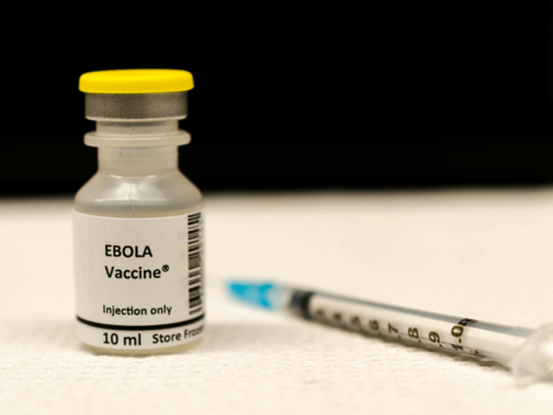 Ebola Vaacine Ebola Virus Disease (EVD) is a highly infectious and often deadly disease caused by the Ebola virus. The virus was first identified in 1976 during an outbreak in Sudan and the Democratic Republic of the Congo. Since then, several outbreaks of EVD have occurred in Central and West Africa, with the largest outbreak to date occurring between 2014 and 2016 in West Africa, which resulted in over 11,000 deaths. One of the most effective ways to control and prevent the spread of EVD is through the use of vaccines. An Ebola Vaccine is a type of vaccine that provides protection against the Ebola virus. There are currently two types of Ebola vaccines that have been approved for use: the rVSV-ZEBOV vaccine and the Ad26.ZEBOV/MVA-BN-Filo vaccine. The rVSV-ZEBOV vaccine was developed by the Public Health Agency of Canada and is licensed to Merck & Co. It is a live, attenuated vaccine that uses a vesicular stomatitis virus (VSV) vector to express the Ebola virus glycoprotein. The Ebola Vaccine works by eliciting an immune response that targets the Ebola virus glycoprotein, which is responsible for the virus's entry into cells. The rVSV-ZEBOV vaccine has been shown to be highly effective in preventing EVD in clinical trials and has been used in several outbreak responses. The Ad26.ZEBOV/MVA-BN-Filo vaccine, also known as the Johnson & Johnson vaccine, is a two-dose vaccine that uses a combination of adenovirus and modified vaccinia virus Ankara (MVA) vectors to express the Ebola virus glycoprotein. The first dose is administered using the Ad26 vector, while the second dose is administered using the MVA vector. The Ebola Vaccine works by eliciting an immune response that targets the Ebola virus glycoprotein, similar to the rVSV-ZEBOV vaccine. The Ad26.ZEBOV/MVA-BN-Filo vaccine has also been shown to be highly effective in preventing EVD in clinical trials and has been used in several outbreak responses. Both the rVSV-ZEBOV and Ad26.ZEBOV/MVA-BN-Filo vaccines have been shown to be safe and effective in preventing EVD. However, there are some differences between the two vaccines that should be considered when deciding which vaccine to use. For example, the rVSV-ZEBOV vaccine requires only one dose, while the Ad26.ZEBOV/MVA-BN-Filo vaccine requires two doses. The rVSV-ZEBOV vaccine has been associated with a higher incidence of side effects, such as fever, headache, and muscle aches, compared to the Ad26.ZEBOV/MVA-BN-Filo vaccine. Additionally, the Ad26.ZEBOV/MVA-BN-Filo vaccine has been shown to provide long-term immunity, while the duration of immunity provided by the rVSV-ZEBOV vaccine is still being studied. Despite the effectiveness of Ebola Vaccine, there are still challenges to their deployment in affected areas. One of the biggest challenges is the need for cold chain storage, as both vaccines require storage at temperatures below freezing. This can be difficult to achieve in areas with limited resources and infrastructure. Additionally, there may be cultural and social barriers to vaccine uptake, as well as challenges in communicating the benefits of vaccination to affected populations. Ebola Vaccine are an important tool in controlling and preventing the spread of EVD. The rVSV-ZEBOV and Ad26.ZEBOV/MVA-BN-Filo vaccines have both been shown to be safe and effective in preventing EVD and have been used in several outbreak responses. 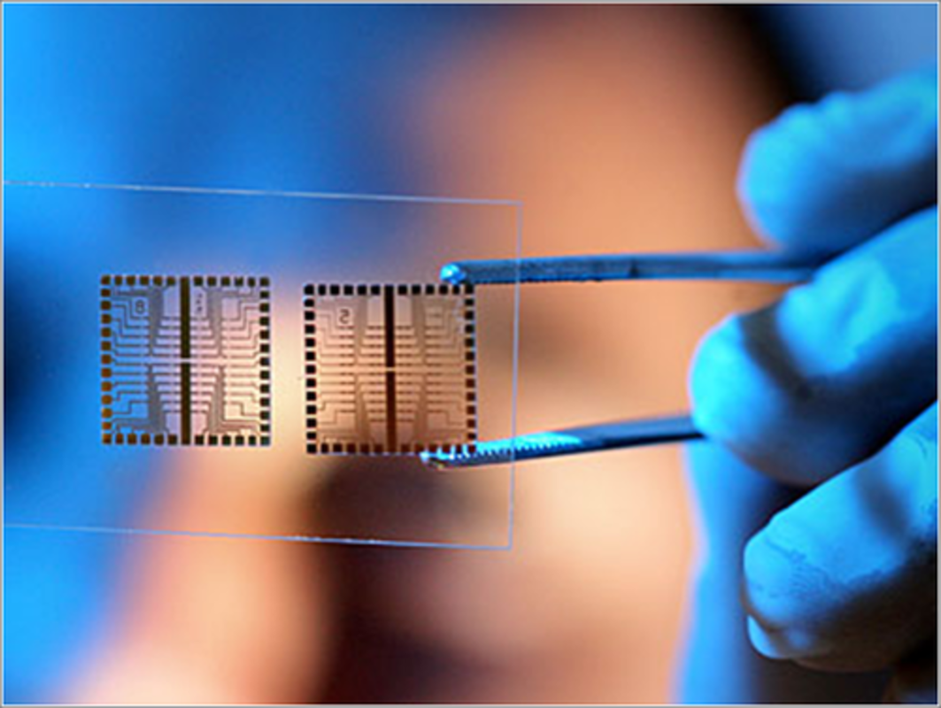 Biochips are small workshops created to function in the living beings. These devices connect semiconductor and organic sciences and allow quick, appropriate tests. They can identify DNA, protein, and other organic molecules. They are utilized in clinical research and medical analysis. The instruments are also utilized in managing the disease and forensic science. The most general kind of a biochip is a laboratory-on-chip. This is a small apparatus comprising millions of small molecular detectors. It can conduct thousands of natural reactions in short span of time. Other kind of a biochip is known as an array. It has tens of millions of small detector materials packed in a small microscopic slide. A sensor scans the pattern of these particle and sends them to a monitor for analysis. These kinds of chips are utilized to distinguish between substitute bases in the SNP site. Additionally, these instruments are suitable to large-scale association survey. Biochips are utilized to translate the arrangements of DNA. Actually, they are an essential part of the technique behind next-cohort sequencing. Few biochip uses comprise populace-based medical survey and medicine screening. Others comprise identifying rare microorganisms. There are several kinds of biochips, comprising passive and active transponders. Biochips are employed in forensic and toxicological analysis. For instance, biochips can be utilized to identify chemical agents in organic warfare. Also, they are utilized in disease regulation and in tagging people. There are two kinds of biochips they are DNA chips and protein chips. Protein biochips utilize a more composite process of surface training. Biochips are small clinical devices that are able to perform several biochemical retorts at once. These instruments are known as micro-reactors as they can conduct thousands of organic reactions in a short span of time. Biochips have various applications in biotechnical research, medical diagnostics, and toxicological survey. For instance, they can be utilized to identify a patient's genetic classification, and identify hazardous biological agents in process. The advantages of biochips comprise their performance, scalability, and quantity. They are vastly efficient and can be utilized for screening several biological analytic, such as proteins, DNA, and insulin. Biochips are vastly acquired in the pharma and biomarker discovery organizations. The techniques have also allowed novel methods for untying the cellular procedure. Additionally, they can be used in NGS, a kind of genetic series. NGS acts as a main feature of technological breakthroughs in drugs. It has enabled for the detection of bacteria, a crucial step in the prior identification of bacterial inflammation. Moreover, it can be utilized for the detection of rare changes. Anyhow, there are considerable limitations linked with NGS. Amongst them, the creation of nucleic acids at a particular concentration is required to perform an analysis. Also, investigative biology-specific method can be difficult. Other major trial of NGS is systematic faults. These errors could cause to imprecise mutation capacities. To address these contests, preprocessing Biochips are accessible to make the nucleic acid library for efficient NGS. These chips are an ensuring methods for the global market. Their benefits comprise their performance, tractability, low budget, and output. 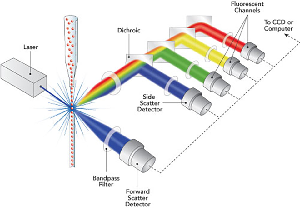 A common cell biology method called Flow Cytometry uses laser-based technology to profile, count, and sort the cells in a heterogeneous fluid mixture. The interaction of cells or other particles suspended in a liquid stream with a laser light beam is monitored by an electronic detecting device as light scatter and fluorescence intensity using a flow cytometer machine. The amount of a cellular component will ideally be represented by the fluorescence intensity if a fluorescent label, or fluorochrome, is precisely and stoichiometrically linked to that component. The automated devices known as Flow Cytometry (FC or FCM) measure the characteristics of one cell at a time. They can quantify a variety of cell properties, including cell size, cell granularity, the quantities of various cell components, including total DNA and recently synthesised DNA, the amount of messenger RNA produced when a gene is expressed, the quantities of particular surface receptors and intracellular proteins, and the quantities of transient signalling events in living cells. When absolute values are required, quantities can be expressed as the number of molecules in a cell. Cell by cell, over 10,000 cells, and up to three to six features or components are often quantitated in a single sample in less than a minute (not counting time to prepare the sample, which might be an hour or more). To distinguish the main groups of leukocytes in peripheral blood, cell size and granularity measurements alone are sufficient. The foundation for clinical equipment that performs automated complete blood counts is this (CBC). Quantitation of particular structures is possible by incorporating fluorescent probes into the cells ("flow cytofluorometry"). The most typical application of Flow Cytometry is for the total DNA per cell in tumour biopsy specimens, for the diagnosis and prognosis of clinical cancer. Quantitation of CD4+ vs. CD8+ T cells in blood is another important application for assessing the effectiveness of anti-HIV medications and determining when an HIV infection has led to AIDS. Research uses Flow Cytometry a lot. In addition to a sizable portion of studies on cell structure, function, and mechanism in other journals, more than one third of papers in the Journal of Immunology also contain flow cytometric data. Monodisperse (single, unclumped) cells are suspended in flow cytometers, where they are passed in single file in front of a laser beam. The amount of dispersed and fluorescent light is measured as each cell moves across the laser beam. Sensitivity is constrained by "autofluorescence," which are naturally fluorescent parts of cells that create a background fluorescence intensity; fluorescent probes must generate much higher intensities for their signals to be reliably quantitated. The majority of Flow Cytometry is analytical; after the data is collected as the material moves through the cytometer, it is discarded. Preparatory flow cytometry sorts living cells into several containers according to their individual characteristics. Fluorescence microscopy (FM) and flow cytofluorometry (FC) can be compared. For a large number of cells, FC can quantify the total levels of a component per cell (typically 10,000, up to 100,000 easily). Normally, FC is unable to determine a component's location within a cell. Flow Cytometry reveals whether a fluorescent component is evenly distributed throughout the cell or concentrated in specific anatomical areas, as well as whether the distribution is time-dependent. The following are the aims of the class's Flow Cytometry experiment:
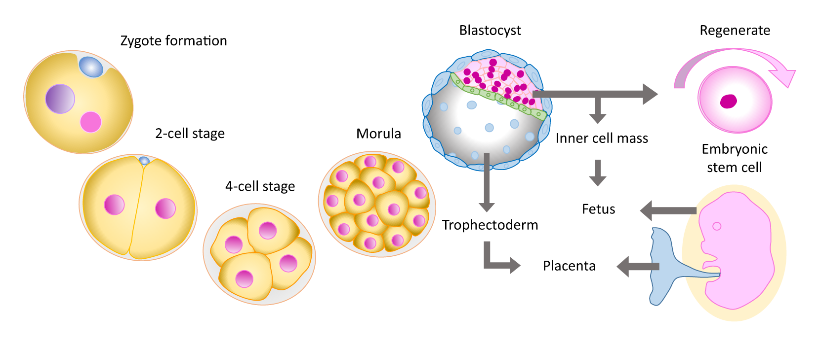 Human Embryonic Stem Cells are the body's raw substances — cells from which all the tissues with particular services are created. Under the proper situations in the body or a lab, Human Embryonic Stem Cells fragment to create more cells known as daughter cells. These daughter cells are either new stem cells or particular cells with a very particular services, such as blood cells, brain tissues, cardiac muscle tissues or bone cells. No other tissues in the body has the direct capability to create new cell forms. Scientists hope Human Embryonic Stem Cells survey can aid to expand knowledge of how disorders take place. By watching Human Embryonic Stem Cells mature into tissues in joints, cardiac muscle, nerves, and other organs and cells, scientists may better know how disorders and situations innovate. Generate healthy cells to replace cells affected by disease. Human Embryonic Stem Cells can be directed into being particular tissues that can be utilized in people to recreate and repair cells that have been destructed or impacted by disorder. People who might be advent from Human Embryonic Stem Cells comprise those with spinal cord wounds, type 1 mellitus, Parkinson's disease, amyotrophic lateral sclerosis, Dementia disease, cardiovascular disorders, stroke, burns, mellitus and osteoarthritis. Stem cells may have the strength to be developed to be new cells for usage in grafting and regenerative drugs. Scientists approach to innovate the knowledge on Human Embryonic Stem Cells and their uses in grafting and regenerative drugs. There are various sources of Human Embryonic Stem Cells. Embryonic stem cells comprises stem cells come from embryos which are 3 to 5 days old. At this phase, an embryo is known as a blastocyst and has around 150 cells. These are pluripotent stem cells, it means that they can fragment into many stem cells or can be of any kind of cell in the body. This adaptability enables Human Embryonic Stem Cells to be utilized to recreate diseased cells and organs. Adult stem cells are those stem cells that are found in small numbers in many adult cells, such as bone marrow. In comparison to human embryonic stem cells, adult stem cells have a very restricted capability to give rise to several cells of the body. Currently, scientists thought adult stem cells can make only same kind of cells. For example, scientists thought that stem cells existing in in the bone marrow could give growth only to blood cells. Anyhow, emerging proofs recommend that human stem cells may be capable to design several kinds of cells. For example, bone marrow stem cells may be capable to make bone or cardiac muscle cells. This scientists has led to initial-stage medical experiments to test practicality and security in people. For instance, adult stem cells are at present being experimented in people with nervous or cardiac disorder. Adult cells changed to have features of Human Embryonic Stem Cells. Researchers have effectively changed normal adult cells into stem cells employing genetic reprogramming. By changing the genes in the adult tissues, scientists can reprogram the tissues to act same as an embryonic stem cells.  Rechargeable batteries, digital cameras, and high-quality image sensors are all built into digital microscopes, and there are also storage choices for photos. There are several different kinds of digital microscopes, including desktop and portable models. Furthermore, not many wireless microscopes have associated display systems. For a Digital Microscope to work, at least one power source is needed. The incorporation of technology into microscopic techniques, like 3D lighting, image-sharing capabilities, and others, eliminates the need for additional instruments to be purchased. Digital Microscopes are classified as Class 1 medical devices by the U.S. Food & Drug Administration (FDA), which means that the restrictions that apply to them are less strict than those that apply to class 2 and class 3 medical devices because they do not affect how the human body functions. Additionally, digital microscopes are categorised by the European Commission using the unified nomenclature that was published in the Official Journal of the European Union. Digital microscopes from the DSX Series, ShuttlePix, and Smartzoom 5 are all offered for purchase. A Digital Microscope is a sophisticated microscope that uses an LED screen in place of the standard compound microscope's ocular. Without using an eyepiece, the image can be seen immediately on the monitor. The microscope's lenses determine how magnified the image will be. The classic or compound microscope can frequently be assisted by a camera that is connected to a computer and has the software necessary to take pictures of the items. These microscopes, however, cannot be classified as digital microscopes. The primary benefit of a digital microscope is the ease and speed with which an object's picture may be viewed while seated comfortably. Additionally, it can be shared and debated with others. When using a microscope for extended periods of time or when a lot of samples need to be inspected, a digital microscope is quite helpful. Desktop Digital Microscopes are largely utilised in forensics, surface metrology, textile manufacturing for inspecting fibre quality, and life sciences research labs. Environmental scientists, horticulturists who need to identify insects and plant diseases, printers in the printing business, and art restorers can all utilise pocket-sized or portable digital microscopes for field work because they are portable and easy to carry. Images taken with wireless digital microscopes can be shared on many screens at once. Digital Microscopes are frequently used in scientific study. It is employed in life sciences to investigate the structure, function, and microstructure of the cell and biomolecules. Finding and examining trace evidence, extracting DNA from blood or hair, and closely scrutinising disputed documents, signatures, handwritings, etc. are all done using it in forensic science. The nanoscale structure of molecules and nanoparticles is studied with a digital microscope in the fields of material science, earth science, and chemical science. In biomedical and pharmaceutical engineering, the digital microscope is essential. Audiologists use it to carefully examine the inner ear, while dentists use it to carefully inspect teeth. The cosmetologist can thoroughly analyse the skin and hair with the use of digital microscopy before continuing with the necessary treatments. Digital microscopes are widely used in industry. It is utilised in metallurgy, medical devices, watches, jewellery, metals and machine engineering, as well as microelectronics and semi-conductors. Molecular Diagnostic is a growing field of research and development, and many different types of tests can be performed to help diagnose a disease or health condition. Some of these tests include PCR, Microarrays, and Single-gene assays. Detecting the micro- and macro-levels of gene expression is an important aspect of modern medicine. Molecular Diagnostics has been applied in many research areas. One of the most important applications of this technology is in the field of drug discovery. With further developments in molecular diagnostics, it is predicted that there will soon be able to better target and deliver our drugs to patients in a more timely fashion. A microarray has been used for many years, but the latest technologies allow for highly multiplexed DNA analysis. One of the most important applications of this kind of technology is to detect the presence of small non-coding RNAs in the genome. Another major application of Molecular Diagnostics is the identification of disease-related genes. These genes are the focus of much of modern biomedical research. This is achieved by comparing and contrasting the gene expression levels of different types of tissue. This can be done by using arrayed primer extension microarrays. Molecular Diagnostics using PCR is an effective method for detecting and diagnosing microbial infections. The method allows the identification of various pathogens with high specificity. It is also beneficial in cases where culture cannot be performed. The method can detect infectious agents that are resistant to conventional methods of culture. PCR is a genetic amplification technique that uses a small amount of DNA. Its benefits include its ability to analyze DNA in a shorter laboratory processing time. It can be used to identify and distinguish infections with chronically persistent agents and to confirm serologic screening tests. It can also be used to identify transmission Cadenas. Molecular Diagnostic testing, or genetic testing, analyzes DNA to identify changes in an individual's DNA that may be associated with a disease or disorder. These tests may identify risk factors for common disorders and may help identify inherited cancer risk. However, genetic testing can also raise new ethical issues. Single gene assays are used in clinical testing to answer clinical questions in high-predictability populations. They are typically performed after a minimally invasive blood draw. These tests can identify specific gene changes in cancer cells. They may also identify changes in drug dosage. For example, a DNA test can identify how a drug interacts with a cellular target. Molecular Diagnostics testing can also be used to assess the impact of genetic diseases on health, including genetic counseling. It can also be used to identify risk factors for common disorders, such as schizophrenia. Single gene assays may be used for testing challenging specimens, such as tumor tissue or liquid biopsy. However, these tests should be used with caution. They may result in an overestimation of disease risk or an increase in patient anxiety. Molecular theranostics is a revolutionary way to treat cancer. They are a promising field that holds tremendous promise for both pharmaceutical and diagnostic companies. Molecular Diagnostics have made their way into all aspects of medicine, including dermatology. These innovations have replaced "excise and pray" approaches with personalized medicine. Molecular theranostics aim to interfere with the growth of cancer during the molecular diagnosis process and ultimately reduce the burden of cancer on patients. Vaccine Adjuvants Are Used To Induce A More Robust Immune Response When Added To Antigens14/11/2022 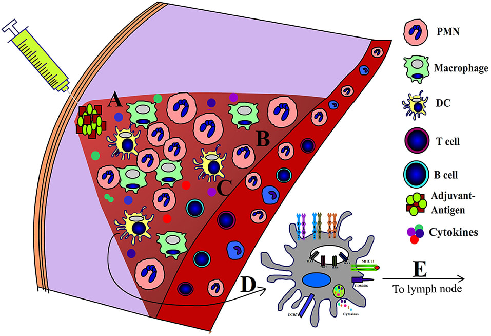 Immunological adjuvants are characterised as molecules, compounds, or macromolecular complexes that enhance the strength and endurance of a particular immune response to antigens, but do so without significantly increasing toxicity or immune effects. The Latin word adjuvare, which means "to help," is used to describe the adjuvant. It works by making the co-administered microorganism, harmless protein, or polysaccharide more immunogenic. The mechanisms of action of adjuvants have long been mysterious. Vaccine Adjuvant are chemicals that increase the strength and longevity of the immune responses triggered by an antigen following vaccination. They consist of both vehicles and immunostimulants, with some of their constituents having both functions. Vehicles- Before administering the vaccine, vehicles of the particulate variety—such as aluminium hydroxide and emulsions—are mixed with the antigen. This causes a "depot effect" (for instance, intramuscular), allowing the antigen to release gradually and to interact with immune cells for a longer period of time. Some vehicles have additional immunostimulatory qualities, such as the ability to activate the inflammasome response when exposed to aluminium hydroxide (alum). Immunostimulants- Adjuvants are chemicals that increase the strength and longevity of the immune responses triggered by an antigen following vaccination. They consist of both vehicles and immunostimulants, with some of their constituents having both functions. Toll-like receptors (TLR), for example, are pattern recognition receptors (PRR) that are known to be activated by immunostimulants. Antigen-presenting cells (APC) can be stimulated by PRR agonists, which enhance antigen presentation and causes the release of pro-inflammatory chemokines and cytokines. In order to maximize the T cell response, the antigen and PRR agonist are combined with the goal of co-delivering both molecules to the same APC. Adjuvants are chemicals that increase the strength and longevity of the immune responses triggered by an antigen following vaccination. They consist of both vehicles and immunostimulants, with some of their constituents having both functions. Antigen-adjuvant conjugates-Contrary to simple combinations of antigen and adjuvant, antigen-adjuvant conjugates are antigen-PRR agonist fusion vaccines that stimulate a more potent immune response. By preventing the two molecules from rapidly dissociating following the delivery of the mixture, this might be explained. Additionally, dendritic cells can internalize conjugated antigens to create an intracellular antigen and immunostimulants depot for persistent T cell stimulation. In terms of scope and speed, the COVID-19 pandemic vaccine development effort is unprecedented. The development of an effective vaccine against SARS-CoV-2 is being pursued using a variety of different approaches, the majority of which call for the addition of an adjuvant to enhance the efficacy and safety of the vaccine. The process of triggering defence mechanisms in fish against pathogenic bacteria by exposing them to non-pathogenic forms or microbe components is known as Fish Vaccines. The three main vaccination delivery methods are intravenously, subcutaneously, and orally. However, only healthy fish should ever receive the vaccination because it is a preventative measure rather than a treatment. Bacterins, live attenuated, subunit vaccines, and toxoids are some of the most frequently used fish vaccine types.  Biotechnology is an expanding scientific field that involves the production of products derived from living organisms. Common applications include biopharmaceuticals, agricultural and food genetically modified crops, and energy production. Agricultural biotechnology is a growing field that can help farmers improve their yields while minimizing harmful impacts on the environment. It can be used to solve a variety of problems, including food insecurity and water scarcity. Agricultural biotechnology includes the process of developing and utilizing genetically modified organisms for a variety of purposes. Biotechnology is an expanding scientific field that involves the production of products derived from living organisms. Common applications include biopharmaceuticals, agricultural and food genetically modified crops, and energy production. Agricultural biotechnology is a growing field that can help farmers improve their yields while minimizing harmful impacts on the environment. It can be used to solve a variety of problems, including food insecurity and water scarcity. Agricultural biotechnology includes the process of developing and utilizing genetically modified organisms for a variety of purposes. Biotechnology can improve the properties of petroleum and coal. It can help reduce the viscosity of oil and cut air pollution. Bioprocessing can also reduce the level of air pollutants. Many potential applications of biotechnology are in energy. Biotech fuels are environment-friendly and do not emit greenhouse gases. Pharmaceuticals can also benefit from biotechnology. In Africa, biotechnology may help fight the problem of hunger and disease. Although there are many uses for biotechnology, the cost remains a major barrier to implementing new technology. Biotechnology has had a dramatic impact on the development of food and agriculture. With its help, scientists are able to create crops with different traits, from improved yields to better taste. For example, scientists have developed a purple tomato that contains a cancer-fighting compound by inserting a bacterial antibiotic resistance gene into the plant's genome. Biotech has also had a profound impact on the field of forensic science. The fields of industrial biotechnology and medical biotechnology have both benefited from the development of biotechnology. These techniques have a wide range of applications in various industries, ranging from the creation of new materials for the construction industry to the production of beer and wine. Industrial biotechnology also has applications in agriculture, as some strains of bacteria have been used in the production of genetically modified crops. Several applications of biotechnology can benefit human health and the environment. These applications include addressing severe acute malnutrition and micronutrient deficiencies. Using microorganisms, biotechnologists are able to address issues like food fortification, probiotics for human health, and detecting GM traits in food. It can also help to solve global challenges such as water and food insecurity. The use of microorganisms in agriculture can reduce pollution in the environment. Biotechnology is used to make life-saving drugs, as it is used in the production of antibiotics and vaccines. Added genetics with desirable traits are introduced into plants to make coded proteins. These products can be used on the body to treat cholera and hepatitis. Biotechnology is also helpful in the production of biofuels. However, the processes involved in these products are complex, so it is important to understand how biotechnology works. Biological processes have been used by humans for tens of thousands of years. About 6,000 years ago, humans began to harness these processes to produce food, alcohol, cheese, and other products. This process has since advanced to the point where it has become a booming industry. Biotechnology is the application of cellular and molecular processes to create novel medicines, foods, and cosmetics. It is also essential for the development of new products. Biotechnology has transformed bacteria into useful human proteins. Those produced by bacteria are then injected into people with mutations, and the results can be used as medicines for specific illnesses. This process has changed the field of medicine and influenced countless fields and industries. With the advent of genetic engineering, mass production of pure and effective biochemicals is now possible. Biotechnology can improve the properties of petroleum and coal. It can help reduce the viscosity of oil and cut air pollution. Bioprocessing can also reduce the level of air pollutants. Many potential applications of biotechnology are in energy. Biotech fuels are environment-friendly and do not emit greenhouse gases. Pharmaceuticals can also benefit from biotechnology. In Africa, biotechnology may help fight the problem of hunger and disease. Although there are many uses for biotechnology, the cost remains a major barrier to implementing new technology. Biotechnology has had a dramatic impact on the development of food and agriculture. With its help, scientists are able to create crops with different traits, from improved yields to better taste. For example, scientists have developed a purple tomato that contains a cancer-fighting compound by inserting a bacterial antibiotic resistance gene into the plant's genome. Biotech has also had a profound impact on the field of forensic science. The fields of industrial biotechnology and medical biotechnology have both benefited from the development of biotechnology. These techniques have a wide range of applications in various industries, ranging from the creation of new materials for the construction industry to the production of beer and wine. Industrial biotechnology also has applications in agriculture, as some strains of bacteria have been used in the production of genetically modified crops. Several applications of biotechnology can benefit human health and the environment. These applications include addressing severe acute malnutrition and micronutrient deficiencies. Using microorganisms, biotechnologists are able to address issues like food fortification, probiotics for human health, and detecting GM traits in food. It can also help to solve global challenges such as water and food insecurity. The use of microorganisms in agriculture can reduce pollution in the environment. Biotechnology is used to make life-saving drugs, as it is used in the production of antibiotics and vaccines. Added genetics with desirable traits are introduced into plants to make coded proteins. These products can be used on the body to treat cholera and hepatitis. Biotechnology is also helpful in the production of biofuels. However, the processes involved in these products are complex, so it is important to understand how biotechnology works. Biological processes have been used by humans for tens of thousands of years. About 6,000 years ago, humans began to harness these processes to produce food, alcohol, cheese, and other products. This process has since advanced to the point where it has become a booming industry. Biotechnology is the application of cellular and molecular processes to create novel medicines, foods, and cosmetics. It is also essential for the development of new products. Biotechnology has transformed bacteria into useful human proteins. Those produced by bacteria are then injected into people with mutations, and the results can be used as medicines for specific illnesses. This process has changed the field of medicine and influenced countless fields and industries. With the advent of genetic engineering, mass production of pure and effective biochemicals is now possible. |
