 Clinical Laboratory Services Clinical Laboratory Services play a vital role in patient care by providing essential diagnostic information that guides medical decision-making. These services encompass a wide range of tests and procedures that analyze bodily fluids, tissues, and other samples to detect, diagnose, and monitor various diseases and conditions. From routine blood tests to complex genetic analyses, clinical laboratory services are indispensable tools in healthcare settings, enabling accurate diagnoses, treatment planning, disease monitoring, and overall patient management. Clinical Laboratory Services serve as the cornerstone of diagnostics, enabling the identification and characterization of diseases. Through various tests, laboratory professionals can detect abnormalities in blood, urine, tissues, and other samples, aiding in the early detection of diseases such as cancer, diabetes, infectious diseases, and genetic disorders. Timely and accurate diagnoses allow for prompt initiation of appropriate treatment strategies, increasing the chances of successful patient outcomes. Moreover, clinical laboratories employ advanced technologies and techniques, such as molecular diagnostics and next-generation sequencing, to identify specific biomarkers and genetic variations associated with certain diseases, providing valuable insights into individualized treatment approaches and targeted therapies. Clinical laboratory services contribute significantly to treatment planning and monitoring by providing healthcare professionals with valuable information about a patient's response to therapy. Regular monitoring of biomarkers and therapeutic drug levels allows for precise adjustments in treatment regimens, ensuring optimal therapeutic efficacy while minimizing potential adverse effects. For instance, in patients undergoing chemotherapy, laboratory tests can assess liver and kidney function, hematological parameters, and tumor markers, helping physicians tailor the dosage and duration of treatment. Additionally, clinical laboratories play a critical role in monitoring chronic conditions such as diabetes, cardiovascular disease, and autoimmune disorders, enabling healthcare providers to assess disease progression, evaluate treatment effectiveness, and implement necessary interventions to manage and control these conditions. Clinical Laboratory Services play a pivotal role in infection control and public health surveillance. By accurately identifying pathogens responsible for infectious diseases, laboratories aid in the prompt implementation of appropriate infection control measures, preventing the spread of infections within healthcare facilities and communities. Additionally, laboratories contribute to disease surveillance efforts by monitoring the prevalence and patterns of infectious diseases, facilitating early detection and response to outbreaks. They also play a crucial role in monitoring antibiotic resistance patterns, helping guide antimicrobial stewardship initiatives and ensuring the appropriate use of antibiotics. Moreover, clinical laboratories are instrumental in conducting tests for notifiable diseases, allowing public health agencies to track and control the spread of communicable diseases and implement targeted interventions to protect population health. Clinical Laboratory Services contribute to medical research and innovation by providing valuable data and samples for scientific studies. Researchers can access de-identified patient samples and health data, allowing them to investigate disease mechanisms, develop new diagnostic tests, and evaluate treatment strategies. Clinical laboratories serve as a rich source of biological specimens, enabling the discovery of novel biomarkers and therapeutic targets that can lead to the development of innovative diagnostic tools and personalized treatment approaches. Furthermore, advancements in laboratory technologies and techniques, such as high-throughput screening methods and genomic sequencing, continue to expand the possibilities for research and contribute to the advancement of precision medicine. Clinical laboratory services are indispensable components of patient care, playing a vital role in disease detection, treatment planning, disease monitoring, infection control, public health surveillance, research, and innovation. Through the analysis of samples and the generation of critical diagnostic information, clinical laboratories enable healthcare professionals to make informed decisions, tailor treatment approaches, and improve patient outcomes.
0 Comments
Robots Used To Perform Disinfection And Cleaning Tasks On Their Own Are Called Disinfectant Robots7/6/2023  Disinfectant Robot In recent years, the world has witnessed a growing concern for cleanliness and hygiene due to the increasing prevalence of contagious diseases. To combat the spread of germs and ensure a safe environment, the utilization of disinfectant robots has emerged as a revolutionary solution. These sophisticated machines are designed to automate and enhance the process of cleaning and sanitization. The Global Disinfectant Robot Market Size Is Estimated To Be Valued At US$ 946.5 Million In 2022 And Is Expected To Exhibit A CAGR Of 35.0% Between 2022 And 2028. Disinfectant Robots are intelligent machines programmed to perform cleaning and disinfection tasks autonomously. Equipped with advanced sensors, cameras, and mapping capabilities, they navigate through indoor spaces with precision. These robots utilize a variety of mechanisms, such as ultraviolet (UV) light, electrostatic sprayers, or chemical disinfectants, to eliminate pathogens and ensure thorough sanitization. One key feature of disinfectant robots is their ability to identify high-touch surfaces, such as doorknobs, elevator buttons, and handrails, which are common breeding grounds for germs. With their advanced algorithms and machine learning capabilities, these robots can efficiently identify and prioritize areas that require intensive cleaning, ensuring comprehensive coverage in a time-efficient manner. Moreover, Disinfectant Robots can be programmed to operate during off-peak hours, allowing them to carry out their tasks without disrupting regular activities. This scheduling flexibility ensures that spaces, such as hospitals, schools, offices, and public transportation, can maintain cleanliness and hygiene standards without causing inconvenience to occupants. The adoption of disinfectant robots brings forth a plethora of benefits in the pursuit of a germ-free environment. Firstly, these robots offer a higher level of accuracy and consistency in disinfection practices compared to manual methods. They can reach inaccessible areas and apply the appropriate amount of disinfectant, leaving no room for human error. By automating the cleaning process, disinfectant robots eliminate inconsistencies and ensure that every surface is treated effectively. Secondly, Disinfectant Robots contribute to the reduction of labor costs and time. In traditional cleaning methods, a significant amount of manpower is required to cover large areas. With the deployment of robots, human resources can be reallocated to more specialized tasks, while the robots handle routine cleaning duties. This streamlining of resources not only optimizes efficiency but also minimizes the risk of cross-contamination caused by human contact with contaminated surfaces. Medical robots are those created specifically for use in specialized medical applications. Medical Robots can carry out a range of medical procedures, including surgery, diagnostic procedures, and patient monitoring. Surgery can be performed by medical robots completely depending on the pre-operative planning of the surgeon. Furthermore, Disinfectant Robots provide a safer alternative for cleaning personnel. By delegating high-risk cleaning activities to robots, workers are protected from potential exposure to harmful pathogens, chemicals, or hazardous environments. This promotes occupational health and safety, fostering a healthier workforce in various industries.  Medical Implants The field of healthcare is currently experiencing a profound transformation, thanks to the remarkable advancements in Medical Implants. These cutting-edge devices are revolutionizing the way we approach patient care, opening up new possibilities and improving the quality of life for countless individuals. As we witness the rise of medical implants, we are entering a new era in healthcare that holds tremendous promise for the future. Medical Implants refer to devices that are surgically placed within or on the body to replace a missing biological structure, support a damaged organ, or enhance bodily functions. They can be made from a variety of materials such as metals, ceramics, or polymers, and are designed to integrate seamlessly with the human body. One of the most remarkable aspects of medical implants is their ability to restore functionality to individuals who have lost it due to injury, disease, or congenital conditions. For instance, cochlear implants have revolutionized the treatment of hearing loss by bypassing damaged parts of the ear and directly stimulating the auditory nerve. This breakthrough technology has allowed countless individuals to regain the gift of hearing, significantly improving their quality of life. Implantable cardioverter-defibrillators (ICDs) are another remarkable example of medical implants. These devices monitor the heart's rhythm and deliver electrical shocks when life-threatening arrhythmias occur, effectively preventing sudden cardiac arrest. With ICDs, individuals with a high risk of cardiac events can live their lives with more confidence, knowing that their implant is constantly monitoring their heart and ready to intervene if needed. In recent years, advancements in Medical Implants have also transformed the field of orthopedics. Joint replacements, such as hip and knee implants, have become increasingly sophisticated and durable, allowing individuals with chronic joint pain or degenerative conditions to regain mobility and lead active lives. These implants are designed to mimic the natural structure and function of the joint, providing a long-lasting solution for patients who would otherwise suffer from severe pain and limited mobility. Moreover, medical implants are also being utilized in the field of neurology. Deep brain stimulation (DBS) implants, for example, have been used to alleviate symptoms of neurological disorders such as Parkinson's disease, essential tremor, and dystonia. By delivering electrical impulses to specific regions of the brain, these implants can help regulate abnormal neural activity and significantly improve the quality of life for patients. In addition to their therapeutic applications, Medical Implants are also being used for diagnostic and monitoring purposes. For instance, implantable glucose monitors provide continuous glucose readings for individuals with diabetes, reducing the need for frequent finger-prick tests. This technology enables better glucose management and empowers patients to make informed decisions about their diet, exercise, and insulin administration. The rise of Medical Implants is not only transforming individual lives but also reshaping the healthcare landscape as a whole. These devices are opening up new avenues for personalized medicine, where treatments can be tailored to the specific needs of each patient. By integrating sensors and connectivity features, medical implants can collect valuable data about a patient's health, allowing healthcare professionals to monitor their condition remotely, detect early warning signs, and intervene promptly when necessary. However, the rapid advancement of medical implants also brings forth challenges that need to be addressed. The safety, efficacy, and long-term performance of these devices must be rigorously evaluated through clinical trials and post-market surveillance. Additionally, issues such as device compatibility, implant rejection, and ethical considerations surrounding the use of medical implants need careful attention. Despite these challenges, the rise of Medical Implants holds great promise for the future of healthcare. As technology continues to evolve, we can expect to see even more innovative applications of these devices, expanding their scope beyond what we can currently imagine. From bioengineered organs to neural implants that enhance cognitive function, the possibilities are truly limitless. 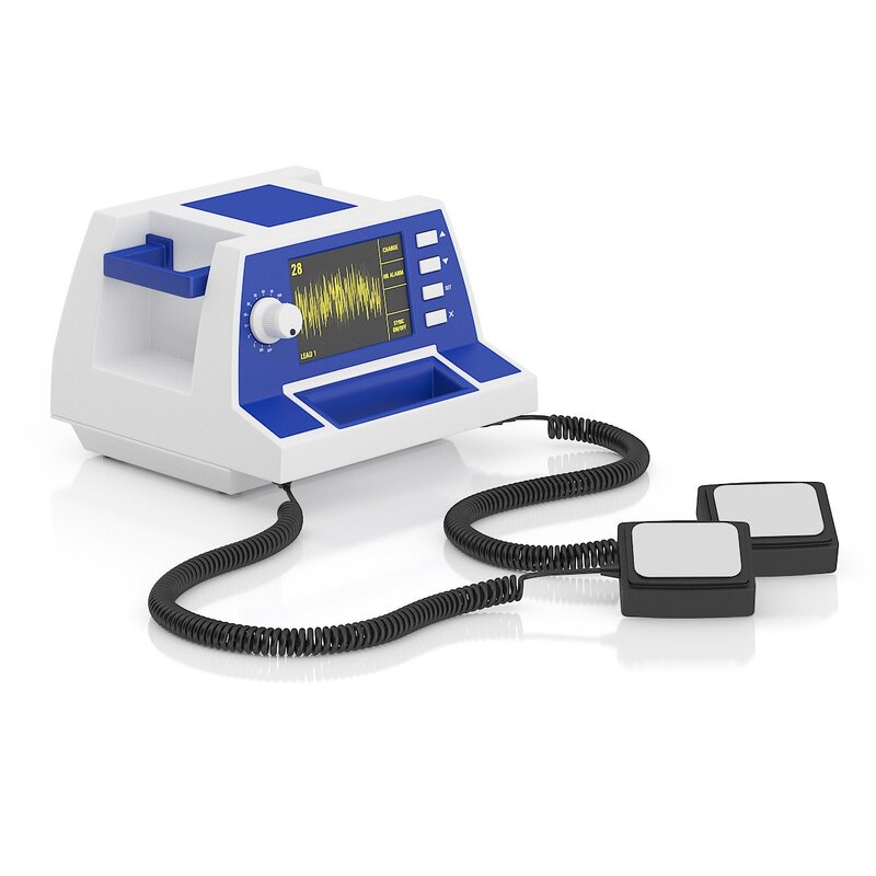 Defibrillators are used in hospitals and other medical settings, as well as public places like airports and schools. They are easy to use by anyone, including people without medical training. Defibrillators can be used to treat life-threatening arrhythmias like ventricular fibrillation (VF) and non-perfusing ventricular tachycardia. Defibrillation sends an electric shock that stops the heartbeat and allows it to beat normally. Defibrillators are most often used in emergency situations when someone goes into cardiac arrest, such as on the job or at public events. Defibrillators can be used by trained health care professionals in hospitals or by laypeople who have been given training and a defibrillator, such as the automated external defibrillator (AED) found in many places today. These devices have two electrode pads that are placed on the chest, one below the right shoulder and the other below the left nipple. These pads have conducting material in them to prevent burns and must be applied with a special kind of wet or solid gel. Once the pads are connected to the defibrillator, the machine checks the person's heartbeat and delivers an electric shock if needed. Defibrillators are the only way to help people survive cardiac arrest. For every minute that passes before a defibrillator is found and delivered, chances of survival diminish. A Defibrillators delivers a shock to the heart, stopping an irregular or life-threatening heart rhythm and restoring it to its normal rhythm. Defibrillation is used to treat dangerous arrhythmias such as non-perfusing ventricular tachycardia or ventricular fibrillation. The automated external defibrillator (AED) in hospitals is a device that prevents sudden cardiac death by treating dangerous heart rhythms. AEDs are designed for use by the general public, and they come with clear instructions for operation. An implantable cardioverter-defibrillator has computer cirduitry, a battery, and wires called leads that connect the generator to the heart. The leads are attached to electrodes that detect a life-threatening arrhythmia and deliver an electric shock to the chest. Generally, the first shock uses a lower energy level and increases in intensity over time. Defibrillators can deliver a high-energy electric shock to help restore a normal heart rhythm. They are used to treat life-threatening arrhythmias that can lead to sudden cardiac death, which occurs when the heart stops beating suddenly and completely. During defibrillation, the device analyzes the heart rhythm through adhesive electrode pads attached to the chest. It advises whether a shock is needed and delivers the electrical current through the pads, causing depolarization of the heart muscles and re-establishing a normal heartbeat. Defibrillators have different electrical waveforms to treat specific conditions. Early paddles required the application of a wet gel to ensure a good connection and reduce the resistance, or chest impedance that would otherwise burn the patient during defibrillation. Later devices were made with self-adhesive electrodes that do not require the need for wet gel. Defibrillators are available in implantable cardioverter defibrillators (ICDs), which are surgically placed inside the body, and wearable cardioverter defibrillators, which rest on the surface of the skin. For people at a high risk of sudden cardiac death, these devices can save lives. Defibrillators can be used to treat a life-threatening heart rhythm called ventricular fibrillation. They deliver a shock using sticky electrodes, often known as paddles, which are placed on the chest. This stops the arrhythmia, and sometimes restores a normal heartbeat. Until recently, the type of electric current delivered through paddle electrodes was only one-way (uniphasic). Biphasic defibrillation is now common and decreases the energy needed to successfully defibrillate, and therefore reduces the risk of damage to the heart. 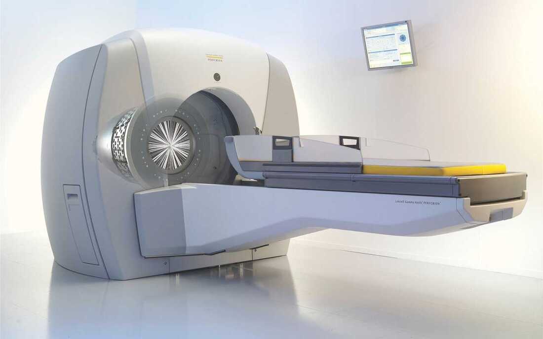 Gamma Knife radiosurgery is a cutting-edge technology that revolutionizes the treatment of brain disorders and tumors. Contrary to its name, it does not involve any surgical incisions. Instead, this advanced medical procedure utilizes highly focused radiation beams to target and eradicate abnormalities in the brain. Gamma Knife radiosurgery has emerged as a precise and non-invasive alternative to traditional surgical interventions, offering patients a safer and more efficient treatment option. Gamma Knife radiosurgery utilizes a unique combination of three-dimensional imaging, computerized treatment planning, and highly focused gamma radiation to deliver precise and targeted treatment to the brain. The system consists of numerous converging beams of radiation, each with a low intensity. Individually, these beams are harmless, but when they intersect at a predetermined target within the brain, they converge to deliver a high dose of radiation, effectively treating the abnormality. The key principle behind Gamma Knife radiosurgery lies in its ability to spare healthy tissues surrounding the target area. This precision is achieved through the use of collimators, which shape the radiation beams, allowing them to conform tightly to the target while minimizing exposure to nearby healthy tissue. The result is an incredibly accurate and efficient treatment that maximizes therapeutic effects while minimizing damage to healthy brain structures. Gamma Knife radiosurgery has found applications in the treatment of various brain disorders, including both benign and malignant tumors, arteriovenous malformations (AVMs), trigeminal neuralgia, and functional abnormalities. It is particularly effective for small to medium-sized tumors, deep-seated lesions, and those located in critical or sensitive areas where conventional surgery carries higher risks. For brain tumors, Gamma Knife radiosurgery offers an alternative to open surgery, enabling the destruction of tumor cells without the need for incisions. This non-invasive approach is advantageous for patients with tumors that are difficult to access surgically or for those who are not suitable candidates for traditional surgery due to age or underlying health conditions. In the case of AVMs, Gamma Knife radiosurgery precisely targets the abnormal blood vessels, causing them to close off over time, reducing the risk of bleeding. Similarly, it has shown success in relieving pain associated with trigeminal neuralgia, a condition characterized by intense facial pain. Gamma Knife radiosurgery offers several benefits compared to traditional surgery and other radiation therapy options. Firstly, it is a non-invasive procedure, eliminating the need for incisions and reducing the associated risks, such as infection and prolonged recovery time. Patients typically experience minimal discomfort and can resume their daily activities shortly after the procedure. Another significant advantage is the precision and accuracy of Gamma Knife radiosurgery. It enables the delivery of a high dose of radiation to the target area while minimizing exposure to healthy tissues, thus reducing the risk of complications and preserving brain function. Additionally, the treatment duration is relatively short, typically lasting from a few minutes to a few hours, depending on the complexity of the case. This allows for a convenient and efficient treatment process, reducing the need for lengthy hospital stays. However, it is important to consider that Gamma Knife radiosurgery may not be suitable for all patients or all types of brain abnormalities. The size, location, and characteristics of the abnormality, as well as the overall health of the patient, need to be taken into account during the evaluation process. In some cases, a combination of treatment modalities, such as surgery and radiation therapy, may be recommended. Furthermore, while the procedure itself is painless, patients may experience some side effects in the days or weeks following Gamma Knife radiosurgery. These side effects can include fatigue, headache, swelling, and temporary hair loss, among others. However, these effects are usually temporary and subside over time. You can Read the Press Release of Gamma Knife 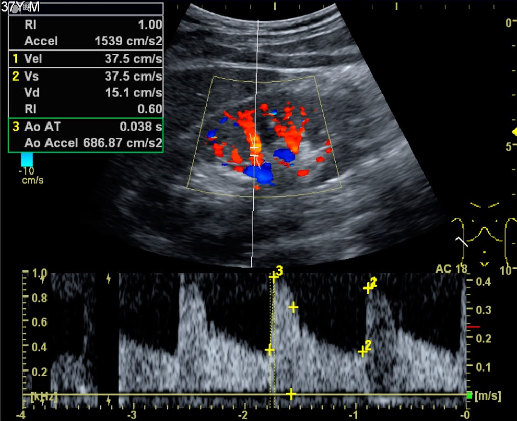 Doppler Ultrasound Doppler Ultrasound is a powerful diagnostic tool that utilizes sound waves to assess blood flow within the body. It provides valuable information about the direction, velocity, and characteristics of blood flow, enabling healthcare professionals to evaluate the circulatory system and detect various abnormalities. The interpretation of Doppler ultrasound requires a combination of technical proficiency, anatomical knowledge, and clinical experience. It is an art that requires a deep understanding of the principles underlying Doppler ultrasound and the ability to integrate that knowledge with patient-specific factors. In this article, we will explore the art of Doppler ultrasound interpretation. Doppler Ultrasound works based on the principle of the Doppler effect, which states that the frequency of sound waves changes when an object is moving towards or away from the source of the waves. In the case of blood flow, the ultrasound machine emits sound waves that bounce off red blood cells and return to the probe. By analyzing the frequency shift of the returning waves, the Doppler ultrasound can determine the speed and direction of blood flow. To effectively interpret Doppler ultrasound, one must first grasp the technical aspects of the procedure. This involves understanding the different Doppler modes, such as color Doppler, spectral Doppler, and power Doppler. Color Doppler provides a visual representation of blood flow direction and velocity, with different colors indicating flow towards or away from the probe. Spectral Doppler, on the other hand, displays the velocity of blood flow over time, represented as a waveform. Power Doppler is sensitive to low flow velocities and is often used to assess small vessels or low-flow states. In addition to understanding the technical aspects, a thorough knowledge of anatomy is crucial for accurate interpretation. Doppler Ultrasound is performed in various areas of the body, including the heart, blood vessels, and organs. Familiarity with the normal vascular anatomy, including vessel course, branching patterns, and anatomical variants, is essential. Furthermore, understanding the hemodynamics of different vessels, such as arteries, veins, and capillaries, is vital to differentiate between normal and abnormal flow patterns. The art of Doppler ultrasound interpretation also requires clinical experience to correlate the imaging findings with patient-specific information. Each patient presents with a unique clinical scenario, and the interpreter must consider the patient's symptoms, medical history, and other diagnostic test results. For example, in the case of a patient with leg pain, the Doppler Ultrasound findings may help determine if the pain is due to arterial insufficiency, venous thrombosis, or musculoskeletal causes. The ability to integrate the ultrasound findings into the broader clinical context is essential for accurate diagnosis and management. Another important aspect of Doppler ultrasound interpretation is recognizing artifacts and differentiating them from true pathological findings. Artifacts are imaging findings that arise from technical limitations or extraneous factors, leading to false or misleading interpretations. Common artifacts in Doppler ultrasound include aliasing, mirror image artifact, and reverberation artifact. Recognizing and minimizing these artifacts requires expertise and can prevent incorrect diagnoses and unnecessary interventions. Moreover, Doppler Ultrasound interpretation involves the application of standardized diagnostic criteria and guidelines. Numerous societies and organizations have established guidelines for the interpretation of Doppler ultrasound in specific clinical scenarios. For example, the Society of Radiologists in Ultrasound (SRU) provides guidelines for the evaluation of deep vein thrombosis (DVT), carotid artery stenosis, and other conditions. Adhering to these guidelines ensures consistency in interpretation and facilitates communication among healthcare professionals. 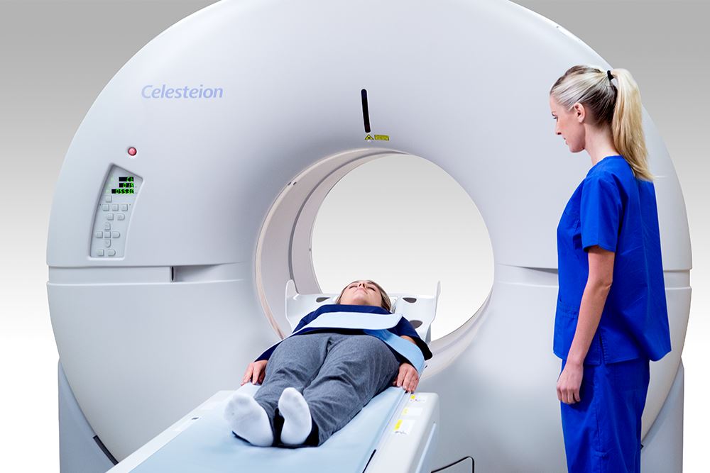 Neurological disorders encompass a broad spectrum of conditions that affect the brain, spinal cord, and peripheral nerves, leading to a wide range of symptoms and impairments. Understanding the underlying mechanisms and identifying early diagnostic markers are crucial for effective management and treatment of these disorders. In recent years, Positron Emission Tomography (PET) Scanners have emerged as powerful neuroimaging tools that provide unique insights into the functioning and pathology of the brain. PET scanners are advanced medical imaging devices that utilize positron-emitting radiotracers to visualize and measure physiological processes in the body. These scanners operate on the principle of detecting gamma rays emitted from the patient's body after the administration of a radiotracer. The radiotracer, typically a compound labeled with a positron-emitting isotope, is injected into the patient's bloodstream and accumulates in the target tissue or organ. As the radiotracer undergoes decay, it emits positrons that annihilate with electrons, resulting in the emission of gamma rays that can be detected by the Positron Emission Tomography (PET) Scanners. The collected data is then reconstructed to create three-dimensional images that represent the functional and molecular activity within the body. Neuroimaging techniques, including PET scanners, have revolutionized our understanding of the brain and its disorders. PET scans provide valuable information about brain metabolism, blood flow, oxygen utilization, and receptor binding. By visualizing these processes, PET scanners enable clinicians and researchers to gain insights into the functioning of the brain in both health and disease. In the context of neurological disorders, PET scans play a crucial role in the diagnosis, staging, and monitoring of various conditions. Positron Emission Tomography (PET) Scanners offer unique advantages in the diagnosis and staging of neurological disorders. In conditions such as Alzheimer's disease, PET scans can detect the characteristic accumulation of amyloid plaques and neurofibrillary tangles in the brain, aiding in early diagnosis and differentiating it from other forms of dementia. Similarly, PET scans can identify hypometabolism patterns in specific brain regions associated with Parkinson's disease, aiding in its diagnosis and monitoring disease progression. PET scans are also instrumental in the evaluation and staging of brain tumors, providing information about tumor metabolism, grade, and response to treatment. The Veterinary Diagnostic Imaging Market was valued at US$ 900.4 million in 2022 and is anticipated to grow at a CAGR of 7.32% from 2023 to 2030 to reach US$ 1578.6 million. Due to the rising zoonotic/chronic disease burden and increasing pet population worldwide, the Veterinary Diagnostic Imaging Market is expanding rapidly on a global scale. Additionally, it is anticipated that increased spending on animal health and rising need for veterinary diagnostics will accelerate market expansion. Positron Emission Tomography (PET) Scanners provide crucial insights into the functional aspects of the brain in neurological disorders. By measuring regional cerebral blood flow and glucose metabolism, PET scans can help identify abnormalities in brain activity. For instance, in epilepsy, PET scans can localize the epileptic focus by identifying regions of abnormal glucose metabolism. In psychiatric disorders such as schizophrenia, PET scans have been used to investigate neurotransmitter receptor function and identify abnormalities in specific brain circuits. Furthermore, PET scans can also aid in understanding the pathology of neurodegenerative disorders by mapping the distribution of abnormal protein aggregates such as tau and alpha-synuclein. By Sending Controlled Electric Pulses To The Heart, Defibrillators Restore Normal Heartbeats22/5/2023 Defibrillators In the realm of emergency medical care, few devices are as crucial and life-saving as Defibrillators. These remarkable machines play a vital role in the resuscitation of individuals experiencing sudden cardiac arrest (SCA), a condition characterized by the abrupt loss of heart function. Defibrillators are designed to deliver an electric shock to the heart, restoring its normal rhythm and potentially saving a person's life. In this article, we will explore the fundamental aspects of defibrillators and delve into their essential role in emergency medical situations. To understand the significance of Defibrillators, it's important to grasp the nature of sudden cardiac arrest. SCA occurs when the heart's electrical system malfunctions, causing the heart to beat irregularly or stop altogether. Within seconds, the lack of blood flow to vital organs, including the brain, can result in unconsciousness, brain damage, or even death. In such critical moments, prompt intervention is paramount, and this is where defibrillators come into play. Defibrillators work by administering an electric shock to the heart, which depolarizes the myocardial cells and allows the heart's natural pacemaker to reestablish its normal rhythm. The electric shock delivered by the defibrillator essentially resets the heart, enabling it to resume its essential pumping function. This process is known as defibrillation and is typically accompanied by cardiopulmonary resuscitation (CPR) to maximize the chances of successful resuscitation. There are two primary types of Defibrillators: automated external defibrillators (AEDs) and implantable cardioverter-defibrillators (ICDs). AEDs are portable devices commonly found in public places, such as airports, schools, and shopping malls. They are designed to be user-friendly and can be operated by individuals with minimal or no medical training. AEDs analyze the heart's rhythm and, if necessary, deliver an electric shock to restore normal heart function. These devices often provide step-by-step voice instructions to guide users through the resuscitation process, making them invaluable in emergencies where immediate medical help may not be available. On the other hand, ICDs are implantable devices that are surgically placed in patients at high risk of SCA. These sophisticated devices continuously monitor the heart's rhythm and can deliver an electric shock if an abnormal rhythm is detected. ICDs are commonly recommended for individuals with a history of heart disease, heart attacks, or other cardiac conditions that increase the likelihood of SCA. By constantly monitoring the heart's activity, ICDs provide a proactive approach to preventing sudden cardiac death and offer reassurance to patients who are at high risk. The importance of Defibrillators lies in their ability to swiftly intervene during a cardiac emergency. Time is of the essence when dealing with SCA, as every passing minute reduces the chances of survival. Research has shown that early defibrillation, within the first few minutes of cardiac arrest, significantly improves the likelihood of successful resuscitation. In fact, the American Heart Association emphasizes the importance of starting CPR and using an AED as soon as possible to improve the chances of survival. Public access to Defibrillators is crucial for maximizing their life-saving potential. This realization has led to the installation of AEDs in various public spaces, creating a more heart-safe environment. Many countries have implemented public-access defibrillation programs, equipping community centers, sports facilities, and other high-traffic areas with AEDs. These programs aim to empower bystanders to take immediate action in case of cardiac emergencies, enabling them to provide critical aid until professional medical help arrives.  A Colposcope gives the doctor extra magnification for viewing the tissues of the vagina and cervix clearly. A doctor uses the colposcope to look at the lining of the vagina and cervix to identify abnormal cells. Using a Colposcope, an Ob/Gyn can closely examine the cervix, vagina, and vulva. It is used if the results of Pap test or an HPV test are abnormal. It can also be used for screening for cervical cancer and to identify precancerous or cancerous cells that may not show up on a Pap test. It is safe, painless and it help the doctor find the right treatment options. It is recommended for women who have had abnormal Pap tests in the past or who are having concerns about their cervix, uterus or other parts of their body. During the exam, a healthcare provider will apply a solution called acetic acid to the cervix and then use a Colposcope to see areas that are abnormal. These areas will stain white, showing if there's anything unusual that needs further study. If the doctor sees something that looks unusual, they may need to do a biopsy. Biopsies help doctors find out if there are abnormal cells on the cervix or vaginal tissue that might lead to cervical cancer. Colposcopy is a common diagnostic procedure used for examining the vagina, vulva, and the cervix. It is usually recommended when a pap smear shows abnormal results or when there is unexplained pelvic pain, inflamed cervix, or bleeding after sexual intercourse. A cervical biopsy may be needed if there are HPV or other conditions that increase the risk of developing cervical cancer, such as unexplained spotting after sex, postmenopausal bleeding, and abnormal results on the Pap test. A doctor or trained nurse uses a special tool called a Colposcope to examine the cervix and vagina to look for 'abnormal cells' that can lead to cervical cancer. These cells are not seen during a normal Pap smear and can be difficult to see using the standard tools the doctor uses for Pap tests. During a colposcopy, the cervix will be lightly swabbed with vinegar or iodine solutions to make it easier for the provider to see any abnormal tissue. If abnormal areas are found, the provider will perform a biopsy. The biopsy involves removing a small sample of tissue to examine under a microscope. The sample will be sent to a lab for further testing. Colposcopy is used to examine the cervix and vaginal walls to check for changes that could lead to cervical cancer. It is a quick and easy way to identify cell changes that can turn into cancer. The procedure is usually safe and does not cause any more pain or discomfort than other speculum exams. A biopsy sample is removed from abnormal areas and sent to a laboratory where it can be checked for signs of pre-cancer or cancer. This biopsy can determine which type of treatment will be needed to remove the cells and prevent them from turning into cancer. A colposcopy is a quick, outpatient exam that allows the health care provider to examine the cervix and vagina.  Dental Floss aids detach wreckage and interproximal dental plaque among teeth. This decreases the threat of gum disorder and tooth decay. Dental Floss is an essential part of any better oral hygiene health and should be utilized regularly. It also aids to detach food elements and bacteria from hard-to-reach regions of the mouth, which can lead to bad breath, craters and gum disorder. The dentist cut off over 18-24 inches of floss and wind it over the middle fingers, leaving over a 1 or 2 inch gap amidst the two. This gives one sufficient length of floss to utilize for all regions in the mouth. One should begin by fixing the Dental Floss amidst the top and bottom teeth and smoothly massage it amidst the teeth utilizing up and down actions. There are several varied kinds of dental floss that are accessible, so one will require to consult to the dentist regarding the kind of treatment that is correct and suitable for the individual. They can also give one suggestion regarding other methods to clean the teeth and decrease the threat of gum disorder. Selecting the correct type of dental floss is important for efficient dental cleaning and will aid one ignore difficulties such as formation of plaque in regions that the toothbrush is incapable of reaching, which can result in gingivitis and periodontal disorders. Unwaxed Dental Floss is created from nylon strands that are turned collectively. It is diluter compared to waxed floss, which states that it will fit in snugger spaces. Anyhow, unwaxed floss is very susceptible to to fraying or breaking. Having a fresh, clean mouth feels better. It also retains the teeth and gums hygienic, which aids one ignore severe difficulties down the road such as tooth decay and gum disorder. The best method to keep the mouth clean is with regular flossing, so one should make it a routine to do so at least two times a day. That way, one can effortlessly get rid of food jiffs and plaque that the toothbrush cannot reach. One can conduct dental floss anytime throughout the day, however one should try to do it prior one brush so the dental floss can aid loosen up food and plaque. One can even utilize a floss pick, which can aid one get into the close-fitting spaces amidst the teeth that routine floss cannot. Dental Floss is created of a variety of substances, consisting plain nylon string and waxed and unwaxed dental floss. Waxed floss is prepared with a light covering of wax to aid it soak substances very efficiently. Flossing is an important part of dental hygiene, however it is not always convenient to keep up with. It is a time-taking task that several people just do not want to do. The best news is that flossing can be a soothing practice that do not have to feel such as a chore. Factually, one can find some great items that make it convenient than ever to floss. One of the most specific benefit to flossing is that it aids inhibit gum disorder, which can lead to very severe health problems later on in life. For instance, it has been associated to strokes and cardiovascular disorders. It can also aid save children from tooth decay and other oral problems by decreasing plaque that may have accrued over the teeth. There are various kinds of Dental Floss that are accessible, and the best one for individual will be based on the particular necessities. One should consult to the dentist or hygienist regarding the kind of treatment that is correct for the individual. One might even find that the dentist will be capable to suggest a brand that will function better for one than what one is using. |
